IT Literature Intelligence 终审版本 (VERIFIED) 论文编号:
307| 原始基线:V0_ZCODE_BASELINE| 语义审核:pass| 图表审核:passAstra裁决:accept_kimi(revise)
The Representation of Tool Use in Humans and Monkeys: Common and Uniquely Human Features
Peeters · Journal of Neuroscience · 2009 · Zotero itemID=2602
这项比较 fMRI 研究让人和猕猴观看同样的动作视频——人手抓物、机械手抓物、以及用螺丝刀、耙子、钳子完成的动作——然后问:理解"使用工具"这件事,在人脑里是否有猴子拿不出来的对应物。答案是"有":两个物种观察手部动作激活了几乎相同的顶叶-前运动网络,但只有人类在左侧下顶叶小叶(inferior parietal lobule, IPL)前部的前缘上回(anterior supramarginal gyrus, aSMG)出现工具交互激活,且这个部位在猴子经过数周真实使用工具训练后依然缺席。对于追踪"哪些认知能力对应哪些新皮层区"这条线的人来说,这是把功能影像与演化问题直接挂钩的一篇代表作。
研究背景
工具使用曾长期被视为人类专属(Oakley, 1956),此后共识修正为:黑猩猩也会自发用工具,而大多数猴类(包括猕猴)只有在训练后才用(Santos et al., 2005, 2006)。争论的焦点因此不是"谁会用工具",而是"谁理解工具与使用结果之间的因果关系"。文献里存在两种对立假说。其一,既然灵长类(无论自发还是训练后用工具)在理解水平上大体相当,那么猴类依靠的是领域一般性的知识,只有人类才拥有专门理解工具功能特性的神经机制,且这一差异应适用于简单与复杂工具(Santos et al., 2006;Johnson-Frey, 2004)。其二,Iriki 等(1996)发现训练用耙的猴子顶叶神经元会获得视-触双模态特性,提示简单工具的使用可能靠两物种共享的机制(Maravita and Iriki, 2004),只有复杂工具才需要人类特有的机制。当时没有人能裁决:到底"用耙子"这类简单工具的神经机制,人猴相似还是不同。
另一个方法是学层面的问题:此前关于工具的脑成像研究几乎全部用静态工具图片(Lewis, 2006 综述),而工具使用本质上是一个动态的、有目标导向的过程;更重要的是,从来没有一项研究让人和猴看同样的动作录像、用同样的对照设计直接比较。这两重缺口正是本文的出发点。
研究思路
作者的整体设计是一条"人找差异、猴验差异"的链条。第一步在人类被试上确立"观察工具动作相对观察手部动作"的交互部位:用 2×2 因子设计(动作类型×动作/静态控制),把"工具动作−静态工具"减去"手部动作−静态手",从而把"看到运动"与"看到物体"从"看到带目标的机械操作"中剥离出来。第二步解决一个混淆:工具动作里的"手"是握着工具的手,与裸手动作的生物性不同——于是加入机械手条件(三指机械臂由隐形的操作者移动、抓握由电脑控制),把"生物手"与"使用器械"两个维度拆开。第三步用三种不同工具(螺丝刀、耙、钳)检验部位的普遍性,并对四组实验的交互做合取(conjunction)分析定出核心区。第四步把完全相同的刺激给未训练猕猴看,用灵敏得多的 MION 对比剂 fMRI 检查猴顶叶是否有人类 aSMG 的对应交互;最后,为排除"猴子不会用工具"这一专业经验的混淆,把两只猴真正训练到熟练使用耙和钳,再扫描一遍。这个阶梯式设计让"阴性结果"也变得可解释:如果连真实使用过工具的猴子都没有,差异就更可能来自物种而非经验。
方法
将“人类被试共 47 名右利手志愿者(平均 23.7 岁),分属 9 组实验”替换为“人类被试共 47 名右利手志愿者(平均 23.7 岁),参与了 4 个主实验(实验 1–4)及 2 个控制实验,部分被试参加多个实验”。:实验 1(n=20,手 vs 机械手)、实验 2(n=21,螺丝刀)、实验 3(n=8,耙)、实验 4(n=8,钳)、两个控制实验(n=6、7),另有 40 人做运动与形状局部izer。刺激为视频(人 13×11.5°,猴 16×13°):一次抓握-拿起动作周期 3.3 s,区组 24 s(猴 36 s)含 7 个(猴 11 个)随机周期;控制条件为静态单帧(实验 2 起每 3.3 s 刷新)与相位打乱视频,另设纯注视基线;控制实验 2 还加入"平移滑行控制"(静态帧整体滑动)与"无目标物体"条件。所有实验要求中央注视,60 Hz 眼动监控,眼跳率人 8–12 次/分、猴 7–20 次/分,条件间无显著差异并作为协变量纳入模型。人类用 3T Philips Intera 采集 BOLD(TR 3000 ms,2.5 mm 层厚,50 层全脑);猕猴用 1.5T Sonata,扫描前静脉注射单晶氧化铁纳米颗粒对比剂 MION/Sinerem(4–11 mg/kg,信噪比约提升 5 倍,且只依赖血容量变化)。分析在 SPM2/SPM5 上做一般线性模型:交互阈值为 p<0.001 未校正,单效应(动作−静态)为 p<0.05 FWE 校正;小样本实验用固定效应、前两个实验用随机效应,最后把四个实验的交互合取。数据投影到人类 PALS 图谱与猕猴 M12 模板(Caret 软件)平面上。猕猴顶叶按细胞构筑划四个 ROI——PF、PFG、PG、Opt(Gregoriou et al., 2006;镜像神经元最密集的 PFG 为重点)——并计算交互幅度(工具动作−静态工具−手部动作+静态手)配两个配对 t 检验(交互检验与动作直接比较)。实验 9 中,两只猴(M13、M14)用行为链塑形法逐步训练(触碰工具→提起→伸向食物→取回食物)使用 25 cm 长的自制耙(3–4 周)与钳(4–6 周,间隔 1 月),随后以标准试次(5 个食物方位)与旋转工具试次考核成绩,并用摄像与标记点做二维轨迹/速度的运动学分析。
主要结果
- 人猴共享"抓取观察"网络。人类观察手部动作(相对静态控制)双侧激活枕颞区、顶内沟前部及邻近 IPL、腹侧前运动皮层加相邻额下回和小脑(图 1A;表 1,如左 DIPSM t=8.50、左 DIPSA t=8.24);机械手条件激活的回路与之相似(图 1B)。未训练猕猴的对应激活(图 7)同样覆盖枕颞、顶叶、前运动与额下回,顶叶激活集中在顶内沟外侧岸,最强处在形状敏感的 LIP 前部、延伸入后 AIP 与后 LIP,且左强于右——两物种在"手怎么抓"层面组织相似。
- 人类出现左 aSMG 工具交互。四个实验(机械手、螺丝刀、耙、钳)都在左侧 IPL 前腹侧凸面、延伸入中央后沟后岸的位置出现"工具动作 vs 手部动作(各相对其静态控制)"的显著交互(图 1C、2;表 2、3);四交互合取给出 75 个体素构成的 aSMG 核心(MNI 峰值 −60,−21,31),59% 落在细胞构筑区 PFt(Caspers et al., 2006)内(图 2E、3B;表 4)。活动曲线证实交互源于动作条件本身的差异而非控制条件的差异(实验 1 双检验 p<0.0001,实验 2 p<0.001,实验 3 p<0.01,实验 4 p<0.05,图 3C–F),且螺丝刀的交互幅度介于耙与钳之间,排除了"只在罕见用法时激活"的解释。
- 交互部位是功能性的、非低层伪影。右半球与左 aSMG 对称的 ROI 从未出现显著交互(图 4);左视野镜像呈现不改变左 aSMG 交互(图 5A,两检验均 p<0.05);交互在平移滑行控制下依然显著(p<0.01),但在视频去掉目标物体后消失(图 5B)——它编码的是带目标的器械操作。枕下回(IOG)的交互部位则完全是视觉性质(形状与运动局部izer 均强,表 4),三工具共同部位位于机械手部位前方,与 aSMG 性质截然不同(图 6)。
- 猕猴顶叶没有对应物。五只未训练猴中,IPL 四区(PF、PFG、PG、Opt)在观察手部或机械手动作时均无显著激活(图 7);tool vs hand 交互只见于视觉区,以 TEO 为主、有时延伸入 STS 或 V4(图 8、9)。对左 PF/PFG/PG 的 ROI 分析中,33 个交互仅 2 个显著(M14 的 PFG-耙、M13 的 PG-钳),接近 1/20 的假阳性率(图 10)。反过来,猴 TEO 的交互模式与人类 IOG 相似(主要源于对静态工具的响应降低,图 11B),说明猴 fMRI 技术本身足够灵敏,阴性结果可信。
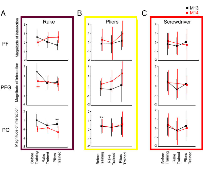
- 真实使用工具的训练也救不回这个部位。M13、M14 用耙成功率达 95%/94%、用钳 97%/76%,工具旋转 90°/180° 后仍能使用(M14 的钳除外),轨迹与速度图极其稳定(图 12)。但训练前后体素水平的交互模式几乎不变,训练后 PF、PFG 对任何工具都无显著交互体素(图 13);ROI 分析中 2×3×3×3=54 次检验仅 3 次显著,仍在机会水平(图 14)。唯一的变化是顶内沟内交互随训练(尤其钳)增强,但弱于人类的 DIPSM——后者是 LIP 前部的可能同源区。
图注解读
图 1 · 实验 1:手部动作观察与机械手交互
原文图注:Figure 1. Results of experiment 1. A, Cortical regions activated (colored red to yellow) by the observation of hand grasping actions relative to their static controls, rendered on lateral views of the standard (MNI) human brain. B, Cortical regions activated in the contrast observation of mechanical hand actions compared with their static control. C, Cortical regions more active during the observation of grasping performed with a mechanical implement than during the observation of the same action by a hand, each relative to its static control. All three contrasts: random effects analysis, n=20, p<0.001 uncorrected. Inclusive masking of the contrasts in A and B with the contrast action observation minus fixation, as used in the monkey (Fig. 7) did not alter the activation pattern. The purple arrow indicates the activation in aSMG. Insets show frames from hand actions and mechanical arm actions.
三行分别是"手−静态手""机械手−静态机械手"与二者之差(交互)的随机效应激活图。A、B 显示抓取观察网络的双侧分布;C 里的紫箭头指向本文的主角——左 aSMG——这是第一次在人类看到"机械器械抓取比生物手抓取更激活"的部位。小图给出两类视频的样帧,帮助读者理解"同一个抓取动作、不同的效应器"的对比逻辑。它支撑结果 1、2。
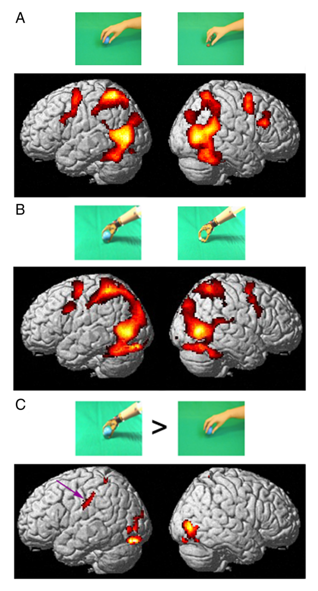
图 2 · 四个实验的左半球交互部位与 aSMG 核心
原文图注:Figure 2. The left hemisphere interaction sites in the four experiments. A, Cortical regions more active (colored red to yellow) during the observation of grasping performed with a mechanical implement than during the observation of the same action done by hand, each relative to its static control, rendered on lateral views of the left hemisphere of standard (MNI) human brain (experiment 1, n=20, random effects analysis). B, Cortical regions active in the contrast observation of screwdriver versus hand actions, relative to their static controls (experiment 2, n=21, random effects analysis). C, SPMs showing significant voxels in the interaction observation of rake vs hand actions (experiment 3, n=8, fixed effects analysis), relative to their static control. D, Same for experiment 4 (observation of actions with pliers, n=8, fixed effects). E, Conjunction of the interactions in the four experiments; threshold for each interaction p<0.005 uncorrected, yielding the core of aSMG. Purple arrows indicate left aSMG.
A–D 用四种不同"工具"重复同一交互设计,左 aSMG 每次都在场——这是"部位普遍性"的直接展示;E 的合取图把四个交互的交集(黑体素)定为核心。读者应注意 A、B 是随机效应、C、D 是固定效应分析,阈值统一在 p<0.001 未校正。它支撑结果 2。
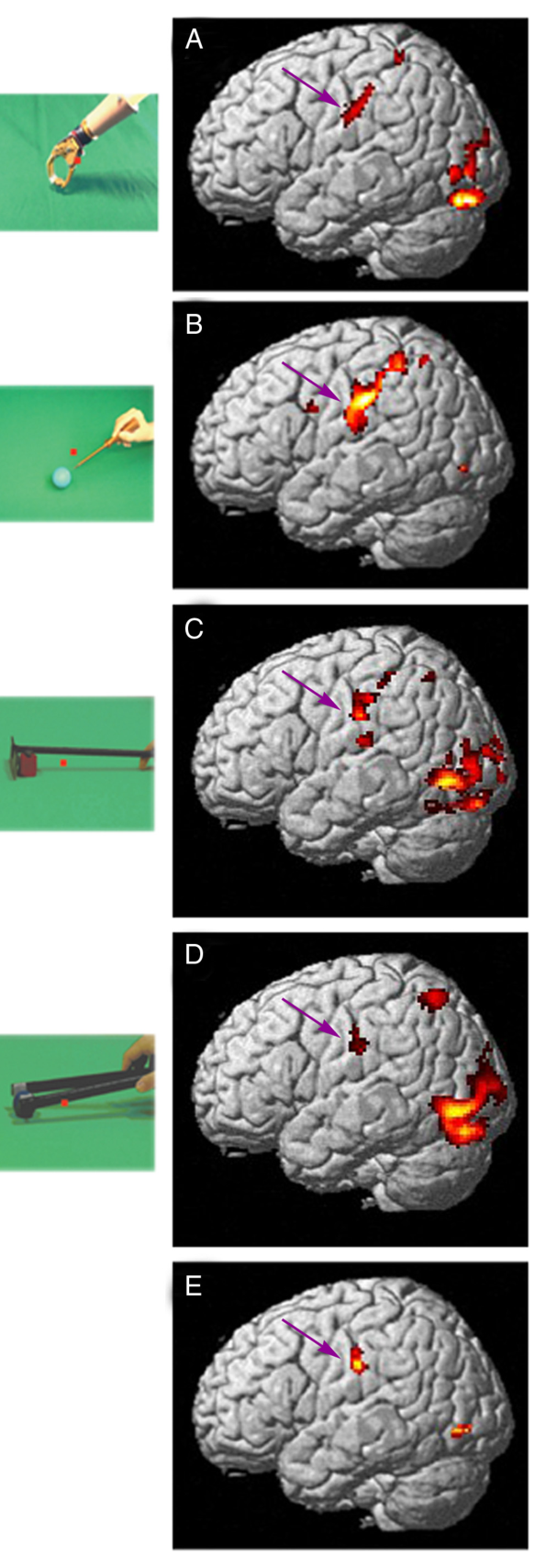
图 3 · 左 aSMG 部位的定位与活动曲线
原文图注:Figure 3. The anterior supramarginal gyrus interaction site. A, Flattened view of the left hemisphere (human PALS atlas). Green areas show activations in the contrast hand action minus static control (p<0.001, experiment 1, same data as Fig. 1A). Blue and red outlines show the regions where the interactions tool versus hand, relative to their controls, were significant (p<0.001) in experiments 1 and 2 respectively (same data as Fig. 2A,B). White lines indicate areas V1-3, yellow dot hMT/V5+. B, Enlarged view of the data shown inside the black square in A. Only interaction sites in the vicinity of anterior SMG are shown. Brown and yellow outlines show the regions where the interactions tool actions versus hand actions, relative to their controls, were significant (p<0.001) in experiments 3 and 4 respectively. Blue and red outlines as in A. Black voxels indicate the conjunction of the four interactions (each p<0.005). Blue ellipses indicate from left to right putative human AIP (phAIP), the dorsal IPS anterior (DIPSA) region and the dorsal IPS medial (DIPSM) region. C–F, Activity profiles of the core of the left aSMG region (black voxels in B) in experiments 1–4. All eight paired t tests were significant…
A、B 把交互部位投到左半球平展图上,与抓取观察激活(绿)及 phAIP、DIPSA、DIPSM(蓝椭圆)对照,读者可以看到 aSMG 核心恰在抓取回路之外偏前;C–F 是四个实验中该核心的活动曲线(相对注视基线的百分比信号变化),工具动作高于手部动作且高于各自的静态控制。C–F 中双配对 t 检验的显著性从 p<0.0001 逐实验降到 p<0.05,对应样本量递减。它支撑结果 2 的定量部分和"aSMG 在手抓取回路之外"的结构描述。
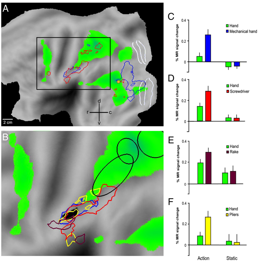
图 4 · 右半球对称 ROI 无交互
原文图注:Figure 4. Right SMG. A, Symmetrical ROI of aSMG core in right hemisphere. B–E, Activity profiles of this ROI in experiments 1–4. In none of the experiments did either paired t test reach significance in this ROI.
在右半球取与左 aSMG 核心镜像对称的 ROI,B–E 显示四个实验里其活动曲线均无交互、动作直接比较也不显著。这是"左半球特异"这一结论的负对照,支撑结果 2 的侧化陈述(也呼应顶叶激活总体左强于右的观察)。
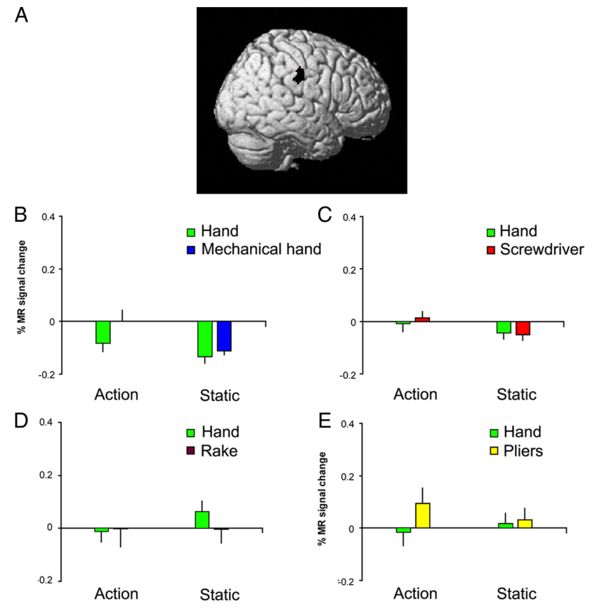
图 5 · 两个控制实验
原文图注:Figure 5. Control experiments. A, Activity profiles of left aSMG in the first control experiment: actions presented in left visual field instead of right visual field (6 subjects); both paired t tests for the interaction and direct comparison of actions (see Materials and Methods) reached p<0.05. B, Activity profiles of left aSMG in the second control experiment: actions and static controls in right visual field with gliding (translation of static frame) controls and tool action with no object added (7 subjects). In both cases the profiles are those of the 5 voxels of the aSMG ROI (used as an a priori prediction) in which the interaction with the screwdriver reached p<0.01 in those particular subjects. In B, the interaction of the tool action with the gliding (translation) and static conditions was significant (paired t test, p<0.01), as was the direct comparison of actions (p<0.001).
A 排除"动作出现在对侧视野"的解释:镜像到左视野后交互依旧;B 排除"平移运动"解释并做关键测试——加入滑行控制后交互仍显著,而去掉目标物体后工具动作信号消失。这两条曲线图共同把 aSMG 的选择性锁定在"对目标物体的器械操作"上,支撑结果 3。
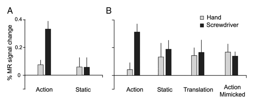
图 6 · 人类 IOG 交互部位
原文图注:Figure 6. Human IOG interaction sites. A, Flatmap of the left hemisphere (human PALS atlas). Green areas show activations in the contrast observation of hand action minus static control (p<0.001, experiment 1). Blue and red outlines show the regions where the interactions tool versus hand, relative to their controls, were significant in experiments 1 and 2 respectively (p<0.001). White lines indicate areas V1-3; yellow dot, hMT/V5+; numbers indicate regions as listed in supplemental Tables S2 and S3, available at www.jneurosci.org as supplemental material; black square, part shown in B. B, C, Part of flatmaps of left and right hemispheres of the human PALS atlas showing the regions corresponding to the conjunction of the interactions for the 3 tools (white voxels), to the interaction for the mechanical hand (blue outlines), and the conjunction of the four interactions (black voxels). These latter voxels, located in IOG, reflect the overlap between two largely distinct regions, probably induced by the smoothing and the averaging across many subjects; green voxels same as in A. D–K, Activity profiles of left (D,F,H,J) and right (E,G,I,K) IOG regions yielded by the conjunction analysis of experiments 1–4 (black voxels in B, C). The larger percentage signal changes in these IOG regions compared with those in IPL (Figs. 3, 5) underscore their visual nature.
这张图提醒读者:合取分析在 IOG 得到的"交集"其实是两个相邻但不同的部位被平滑平均拼在一起的产物——三工具共同部位(白)位于机械手部位(蓝)之前。D–K 的活动曲线显示这些 IOG 区域的百分比信号变化远大于顶叶、且形状/运动局部izer 均敏感,说明它们是纯视觉的刺激差异检测器,与 aSMG 性质不同。它支撑结果 3 的"区分视觉伪影与功能交互"部分。
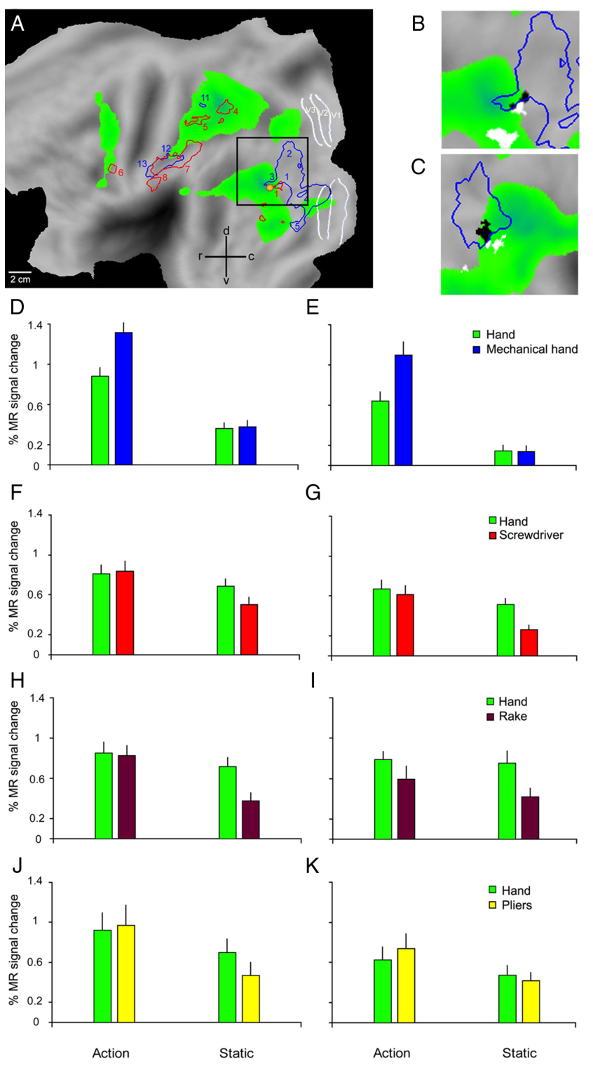
图 7 · 猕猴观察手部与机械手动作的激活(实验 5)
原文图注:Figure 7. Results of experiment 5. A–C, Folded left and right hemispheres (A) and flatmaps (B, C) of left and right hemisphere of monkey template (M12) brain (Caret software) showing cortical regions activated (n=2, fixed effects, p<0.05 corrected, experiment 5) in the subtraction observation of hand action minus static hand (A, B) and observation of mechanical hand action minus static mechanical hand (C). Both subtractions were masked inclusively with the contrast action observation minus fixation. Black outlines, PF, PFG, PG and Opt; outlines of visual regions (V1-3, MT/V5) are also indicated. Color bars indicate T-scores. See also supplemental Figure S1, available at www.jneurosci.org as supplemental material.
用与人类实验 1 完全相同的刺激扫两只猴:A、B 是手部动作激活,C 是机械手动作激活,均以"动作观察−注视"做包容性掩蔽。激活覆盖枕颞、顶叶、前运动/额下回,与人类相似;关键在于黑线勾出的 IPL 四个细胞构筑区(PF、PFG、PG、Opt)内没有激活。它支撑结果 1 的猴侧部分和结果 4 的前提。
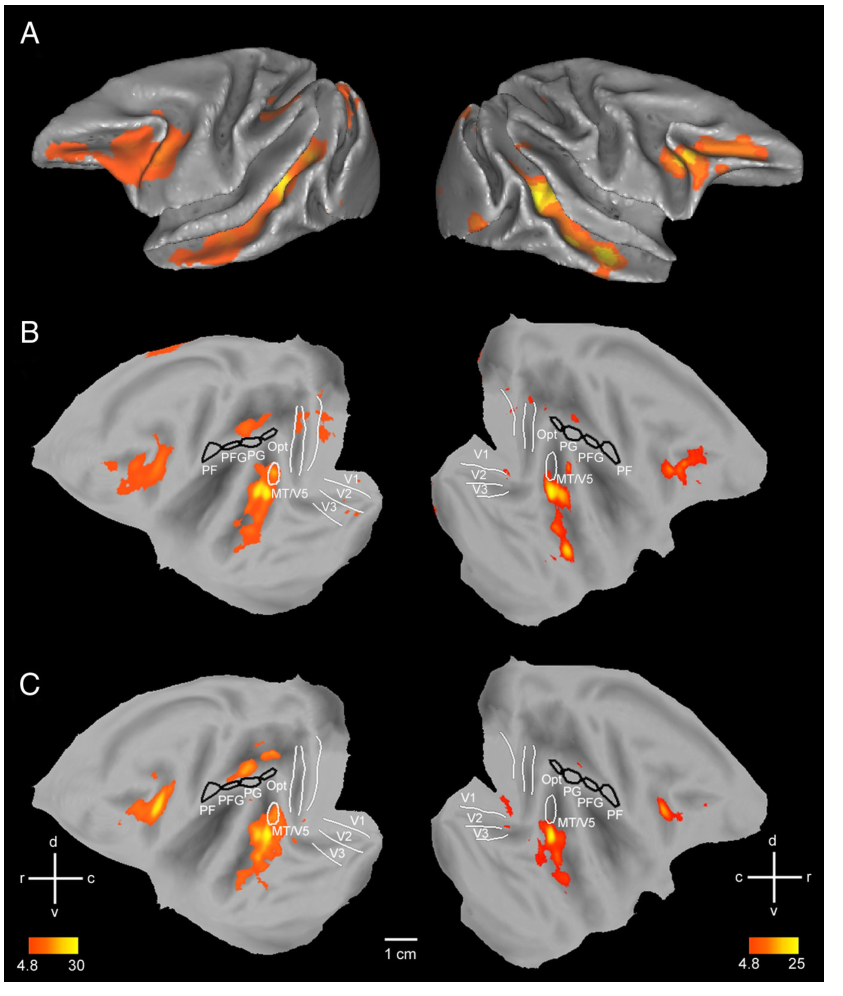
图 8 · 猕猴左半球的工具交互部位
原文图注:Figure 8. Interaction sites in the monkey. A–D, Monkey cortical regions activated during the observation of hand grasping action relative to their static controls (green voxels, fixed effect, p<0.05 corrected, experiment 5, same data as in Fig. 7B). Data plotted on flatmaps of the M12 left hemisphere. The voxels more active (fixed effect, p<0.001) in the contrast tool versus hand actions relative to their static controls (experiments 5–8) are shown in blue (A, mechanical hand), red (B, screwdriver), brown (C, rake), and yellow (D, pliers). ROIs as in Figure 7.
绿色为手部动作观察激活底图,四色分别为机械手、螺丝刀、耙、钳的交互体素。一眼可见:交互体素全部落在枕颞视觉区(TEO 为主)和低级视觉区,IPL 四区内几乎空白(唯一例外是实验 7 耙在左 PG 的少数体素)。它支撑结果 4 的体素水平结论。
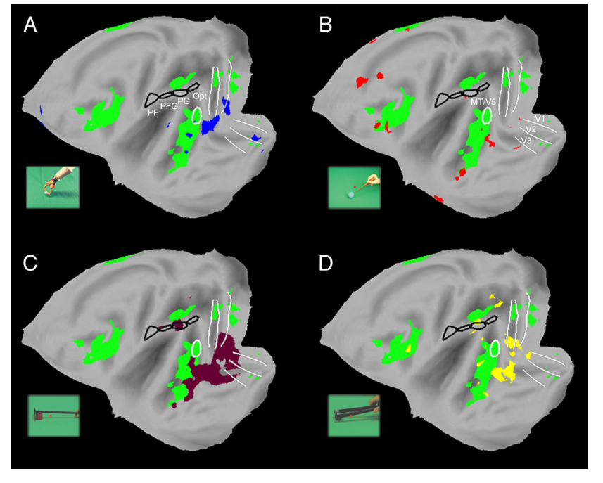
图 9 · 猕猴右半球的交互部位
原文图注:Figure 9. Interaction sites in the monkey. A–D, Flatmaps of the right hemisphere of monkey template (M12) brain (Caret software) showing regions significant (fixed effects, p<0.05 corrected) in the interaction in experiments 5–8 (same color code as in Fig. 8, n=2 in experiments 5 and n=3 in experiments 6–8). ROIs as in Figure 7.
右半球的对应版本,结论与图 8 一致:右 IPL 四区同样没有显著交互。与人类"只有左侧有"形成对照的是,猴两半球顶叶都没有——物种差异不是简单的侧化差异。它支撑结果 4。
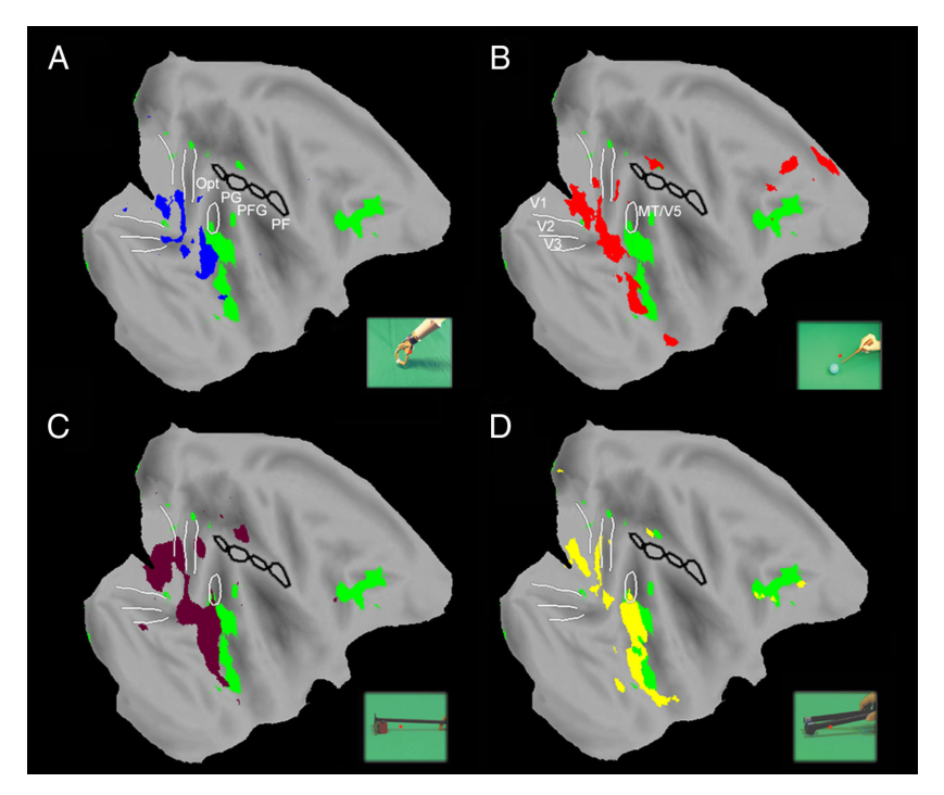
图 10 · 猕猴 IPL 的 ROI 分析
原文图注:Figure 10. ROI analysis of monkey IPL. A, Activity profiles in PFG (ROI analysis, experiment 7). B, Magnitude of the interaction (b − d − a + c) in PF, PFG and PG for different tools and monkeys. Stars indicate significant interactions. Notice that the larger percentage signal change in the monkey reflects the use of contrast agent enhanced fMRI (see Materials and Methods).
A 显示实验 7(耙)中 PFG 的活动曲线:个体间交互方向不一致;B 把每个"工具×猴"组合的交互幅度画在 PF、PFG、PG 三个 ROI 上,星号标显著者。33 个顶叶交互中只有 2 个过双配对 t 检验(约 1/20 的假阳性率预期),且没有系统方向。注意猴的百分比信号变化数值大是因为 MION 对比剂增强。它支撑结果 4 的 ROI 水平结论,是把"体素图阴性"升级为"定量阴性"的一步。
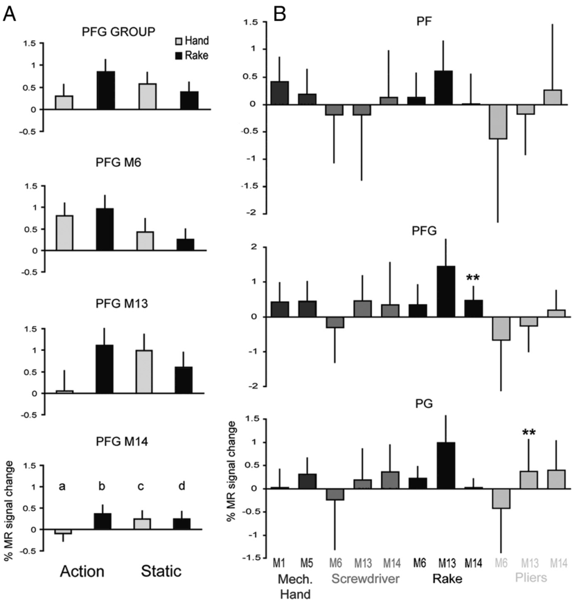
图 11 · 猕猴枕颞交互部位(TEO/V4)
原文图注:Figure 11. Occipitotemporal interaction sites in the monkey. A, Flatmap of left hemisphere of monkey 12 (template brain) showing the regions activated (fixed effect, p<0.001) in the contrast viewing hand action minus viewing static hand (green voxels, experiment 5) and in the interaction for the mechanical hand actions (blue outlines, experiment 5) as well as the voxels common to the interactions for the 3 tools (white voxels, experiments 6–8); ROIs as in Figure 7. B, Activity profiles of the local maximum and 6 surrounding voxels in TEO (white area in A) in experiments 6–8. Again, the larger percentage signal changes in the monkey reflect the use of contrast enhanced fMRI (Denys et al., 2004b) using the V1 activation as common reference estimated the percentage signal changes with MION to average ≈2.5-fold those obtained with BOLD at 3T. A similar factor applies here when comparing to the data of Figure 6, taking into account the larger number of human subjects from whom data are averaged.
A 显示三工具共同交互部位(白)位于机械手交互部位(蓝)前方,主要在 TEO、机械手交互主要在 V4——与人类 IOG 的排列一致;B 显示 TEO 内的交互主要来自对静态工具的响应降低,与人类 IOG 相同。这张图是"猴 fMRI 灵敏度足证"的证据:同类视觉区在人猴身上给出了同类交互。它支撑结果 4 的可信度论证。
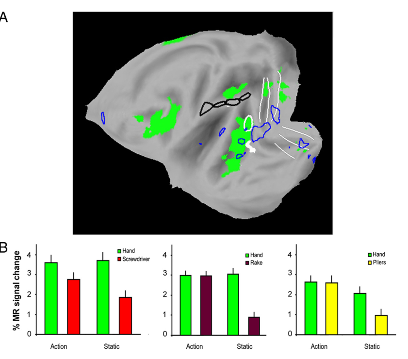
图 12 · 训练后猴使用工具的运动学分析
原文图注:Figure 12. Effects of training in M13 and M14: kinematic analysis. A, C, E, G, Tool trajectories (mean ± 1 SD) for reaching and retrieving food of trained monkeys. Black squares: starting points. Colored squares: food position (5 directions: −45 to +45°). A, E, Trajectories using rake M13 and M14. A marker was attached at the middle of the rake-head. The left starting point was used for all directions except that straight ahead. C, G, Trajectories using pliers (M14). Markers attached on the arms of the pliers, 3 cm from the tip. B, D, F, H, Mean (±1 SD) speed-time diagrams for two trajectories (−2.5° and +45°). Rake, M13 and M14 (B, F); pliers, M13 and M14 (D, H).
A/C/E/G 画出耙头或钳尖标志点的轨迹(含 SD 带),B/D/F/H 是两个代表性方位的速度-时间图。轨迹极稳定、速度模式一致(远端食物先快后慢的拉拽相位),证明两只猴确实"熟练"而非碰巧会用;M13 动作更慢更稳、M14 略快。这张图为"训练有效"提供了行为学证据,是结果 5 的前提。
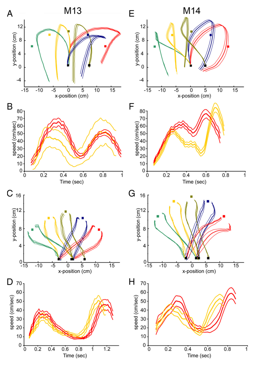
图 13 · 训练前后体素水平的交互变化
原文图注:Figure 13. Effects of training: voxel-based analysis. A–C, SPMs showing significant interaction (n=2, fixed effects, p<0.001) tool action versus hand action observation, relative to their static control, for rake (A), pliers (B) and screwdriver (C) observation before training, after rake training, and after pliers training. The significant voxels (orange-yellow) are overlaid on flatmaps of the left hemisphere of monkey M12.
三列工具、三行训练阶段(训练前、学会耙后、学会钳后)的交互图并排:变化最明显的是 IPS 内交互随钳训练增强,但 PF/PFG(aSMG 的人类对应候选区)在训练后的任何一行都没有显著交互体素。它支撑结果 5 的"训练不改变结论"部分,也支撑讨论中"交互增强发生在抓取回路之内"的解读。
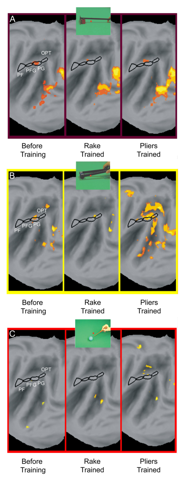
图 14 · 训练后 ROI 交互幅度的纵向分析
原文图注:Figure 14. ROI analysis of the magnitude of the interaction (see Fig. 10 for definition) after training. A–C, Interaction magnitude is plotted as a function of epoch for the two subjects (M13, M14) in each of the three ROIs (PF, PFG, and PG) for rake action observation (A), the pliers action observation (B) and the screwdriver action observation (C). Significant values (both paired t tests p<0.05 corrected for multiple comparisons) are indicated by stars. Runs from A–C were tested interleaved at each epoch.
把每个 ROI 的交互幅度对时间点(epoch)画出来:M13、M14 两条曲线在 PF、PFG、PG 三个区都随训练阶段起伏不定,星号(显著)零星出现——54 次检验中 3 次,符合机会水平。最关键的一行读法:即使看"猴子刚学会用的那个工具",对应 ROI 的交互也不显著。它支撑结果 5 的定量结论。
讨论
作者的结论分两层。功能层面:人猴共享的抓取观察回路把"用工具完成的动作"当作与手抓取等价的动作来处理,而人类额外的左 aSMG 才以"工具的预期用途与使用结果之间的因果关系"为编码维度;aSMG 与以往静态工具图片激活的后部 IPL 不同(后者更可能表征"工具如何被握住",与 Valyear 等 2007 描述的 AIP 后侧工具命名区相区分),也与语言网络无关——它比参与视听语音加工的 SMG 各区更靠前,动作动词激活的岛盖区 SII(Rüschemeyer et al., 2007)则在其前腹侧,而因果知觉的顶叶激活又都位于它更尾部,因此其激活既不能归于词汇加工,也不能归于一般因果推断。结合 SMG 在工具使用想象、哑剧和虚拟工具操作中的激活以及它对工具使用声音的响应(Lewis et al., 2005),作者推测 aSMG 是"观察到的工具使用与工具使用动作程序相匹配"的节点。演化层面:虽然围绕 IPL 同源关系尚有争议(Brodmann 认为人类 IPL 是演化新皮层,而顶内沟本身是古老脑沟),本研究提示 IPL 近顶内沟部分在两物种间功能相似,而 IPL 嘴侧部是人类新皮层区。作者据此把时间线拉到古人类学:奥杜威工业(2.5 Ma)的石片与阿舍利工业(约 1.5 Ma)的手斧多样性暗示因果表征能力,aSMG 的出现"至少可上推至 150 万年前",甚至更早。局限方面作者承认:猴的训练时长远不及人类的终身工具经验,更久更多工具的训练结果未知;由于猴顶内沟区对手与工具动作都有响应,不能排除那里散在着少量工具特异神经元、只是低于 fMRI 的分辨极限(Joly et al., 2009)——这正对应摘要里"tool-specific neurons might reside in the monkey grasping regions"的谨慎表述;训练还可能通过神经连接改变(Hihara et al., 2006)招募新顶叶区,留待后续发表。与此同时,作者也把阴性结果的可信性做足了:5 倍信噪比的对比剂、比全脑分析更灵敏的 ROI 设计、细胞构筑定位、以及 IOG/TEO 交互在人猴间的对称出现。
一句话总结
这篇论文最有力量的地方在于它的阴性结果:一个部位在人身上稳定出现、在猴身上反复缺席,而且缺席发生在猴子真的学会用耙子和钳子之后——物种差异因此很难用经验差异打发。按我的理解,它把"镜像系统之外还有没有人类专属的高阶动作理解区"这个问题落到了解剖上,但"理解因果关系"仍是从激活位置反推的功能标签,aSMG 到底编码因果还是编码工具的运动图式,fMRI 本身无法裁决;这个问题后来也确实是颅外影像与电生理继续缠斗的战场。
审校与证据追溯 (Verification & Evidence)
图表审计结果
- Fig1: 提取质量
good,对齐度full,识别面板[A, B, C] - Fig10: 提取质量
good,对齐度full,识别面板[A, B] - Fig11: 提取质量
good,对齐度full,识别面板[A, B] - Fig12: 提取质量
good,对齐度full,识别面板[A, B, C, D, E, F, G] - Fig13: 提取质量
good,对齐度full,识别面板[A, B, C] - Fig14: 提取质量
good,对齐度full,识别面板[A, B, C] - Fig2: 提取质量
good,对齐度full,识别面板[A, B, C, D] - Fig3: 提取质量
good,对齐度full,识别面板[A, B] - Fig4: 提取质量
good,对齐度full,识别面板[A, E] - Fig5: 提取质量
good,对齐度full,识别面板[A, B] - Fig6: 提取质量
good,对齐度full,识别面板[A, B, C] - Fig7: 提取质量
good,对齐度full,识别面板[A, B, C] - Fig8: 提取质量
good,对齐度full,识别面板[A, B, C, D] - Fig9: 提取质量
good,对齐度full,识别面板[D]